Image Of Spinal levels of the sacral plexus branches mnemonic duration. Learn to draw the brachial plexus in under 4 minutes.

Image Of Color Coding Standards For The 12 Lead Ecg

Image Of Pdf Semi Automated Dti Measurement Of The Brachial Plexus

Image Of How To Study For Anatomy
Key branches of axillary artery.
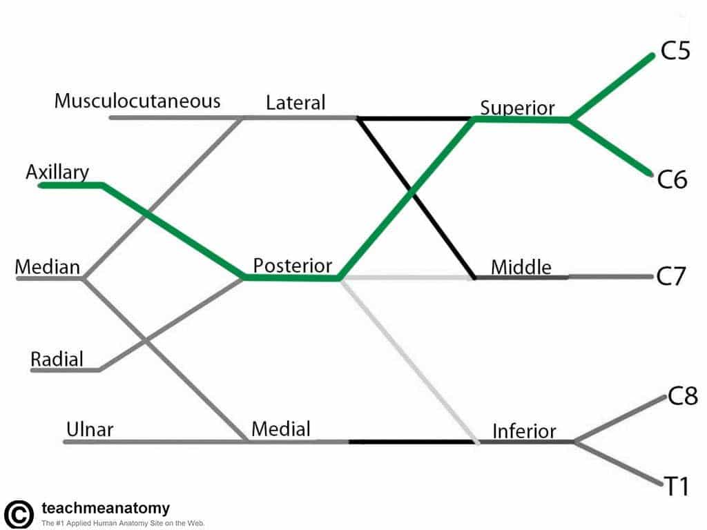
Image Of
Color coded brachial plexus drawing.
The target audience is medical dental pa and pt students of anatomy.
Based on an unpublished illustration by jane phillips conroy for the course principles of human anatomy and development l48 4581 at washington university in st.
The first 3 is the branches to c5 6 and 7 which form ltn long thoracic nerve.
Image Of
It is important to remember that c5 also gives fibres which join fibres from c3 and c4 to form the phrenic nerve.
Structure of the brachial plexus.
Skip navigation sign in.
Image Of
Draw the brachial plexus in 376 seconds or better duration.
More complex diagramming of the brachial plexus includes the four 3s neurosurgeons neurologists and physiatrists will use this diagram system.
Michael dauzvardis 164557 views.
Image Of
Learn how to draw brachial plexus in less than 10 seconds through this video.
This video is unavailable.
The dorsal scapular nerve the long thoracic nerve and the first intercostal nerve.
Image Of
Upper limb sensory and motor examination this card features a dermatomal and peripheral nerve sensation map of the upper limb.
The brachial plexus is formed by the ventral primary rami of the lower four cervical nerves and the first thoracic nerves c5t1.
Be able to draw the brachial plexus in your sleep.
Image Of
Major components robert taylor drinks cold beer.
How to draw the brachial plexus including the 16 peripheral nerves.
There are five nerve roots from c5 t1 which give three nerve branches.
Image Of
Great for mapping out neurological lesions accurately.
Next each of the headless arrows has three nerves attached to it.
1 posterior cord all 3 posterior divisions 2 lateral cord anterior divisions of upper and middle trunks contains fibers for c56 and 7 3 medial cord continuation of the anterior divisions of the lower trunk fibers from c8 and t1.
Image Of
Brachial plexus is the most asked topic in anatomy final exams during first year of mbbs life.
Details on branches from the roots trunks and cords.
It also features myotomes a map of the brachial plexus and some importation conditions.
Image Of
The brachial plexus with the courses of the spinal nerves shown in color.
Add the upper 6 middle 7 and lower subscapular 8 nerves.
Learn to draw the brachial plexus in under 4 minutes.

Image Of Mononeuropathy Disease Malacards Research Articles Drugs
Image Of Search Anatomy Of The Spinal Column Spine Color Coded

Image Of Downloads Study With An Spt

Image Of Brachial Plexus Plexus Products Gross Anatomy Neurology

Image Of Brachial Plexus Color Axillary Nerve Wikipedia The Free

Image Of Brachial Plexus Injury Understanding The Brachial Plexus
Image Of Neurologic And The Cervical Spinal Cord Nerve Plexus And
Image Of Frontiers Rehabilitation Of Upper Extremity Nerve Injuries

Image Of Learning Contract Stephanie Soto Physical Therapy Internship

Image Of Brachial Plexus Drawing

Image Of Quick Draw Anatomy For Anaesthetists Critical Care Northampton

Image Of Brachial Plexus Lesions Drawings Of Explorations And
Image Of Block Diagram Of A Coded Excitation Cfi System In Which

Image Of Norwichra Hashtag On Twitter
Image Of Search Color Coded Skeleton

Image Of Figure 1 From Brachial Plexus Mr Imaging Accuracy And

Image Of Ultrasound Guided Interscalene Brachial Plexus Block Nysora
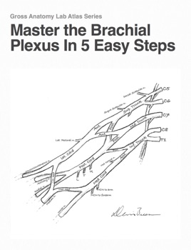
Image Of Master The Brachial Plexus In 5 Easy Steps
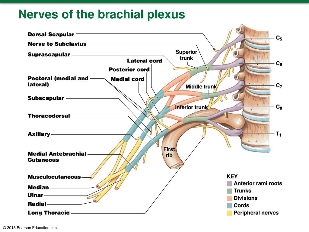
Image Of 2018 Pearson Education Inc Ppt Download

Image Of Pancoast Cancer Tnm Staging Atlas With Oncoanatomy 2e

Image Of Nerve Plexus Wikipedia

Image Of Pancoast Cancer Tnm Staging Atlas With Oncoanatomy 2e

Image Of Three Dimensional Reconstruction Of The Cranial And Anterior

Image Of Everyday Anatomy A Guide Reference Home Study With Bonus 9 Free Ceus

Image Of Amazon Com Practical Management Of Pediatric And Adult

Image Of Dti Tractography Of The Brachial Plexus In A Normal

Image Of Color Coded Vertebral Column Skeleton Anatomy Anatomy

Image Of Ge Signapulse Spring 2018 Imaging The Brachial Plexus In

Image Of Therapy Teaching And Learning Resource Anatomy Club Teaching Templates
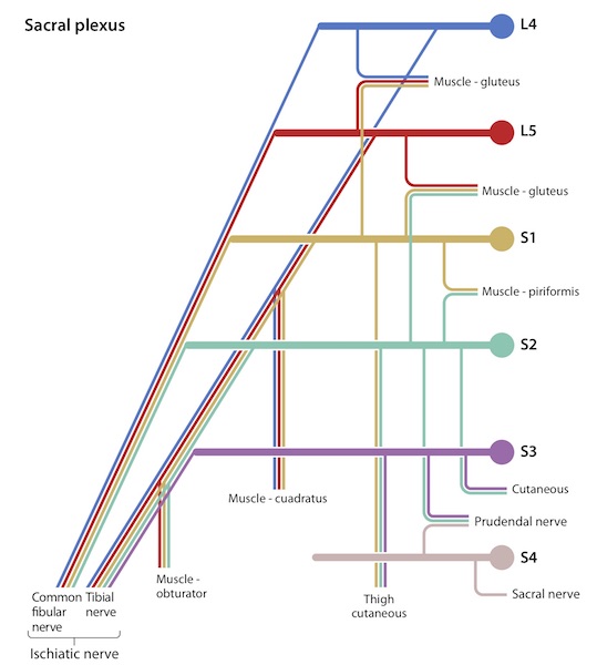
Image Of Anatomy And Physiology In Context Reading Assignment
Image Of The Brachial Plexus Sections Branches Teachmeanatomy

Image Of How To Do 3d Modeling For Ct Surgery Videos Mayo Clinic

Image Of Gross Anatomy Drawing The Lumbosacral Plexus
Image Of Diffusion Tensor Mri Of The Healthy Brachial Plexus

Image Of Figure 5 From Spatial Characterization Of The Motor Neuron
Image Of Diffusion Tensor Mri Of The Healthy Brachial Plexus
Image Of Shoulder Stabilizer Brace Shoulder Rehabilitation Support

Image Of Quick Draw Anatomy For Anaesthetists Critical Care Northampton
No comments:
Post a Comment